Nicholas Whipple

About
Intermountain Primary Children's Hospital Neurosurgery Department and The Sorenson Center for Cancer & Blood Disorders
Nicholas Whipple, MD, MPH, received his medical degree from the University of Mississippi School of Medicine in Jackson, MS. After completing his pediatric residency at the University of Utah and Primary Children's Hospital in Salt Lake City, UT, Dr. Whipple completed 3 years of fellowship training in Pediatric Hematology-Oncology and 1 year of Fellowship training in Pediatric Neuro-oncology at St. Jude Children's Research Hospital in Memphis, TN. During fellowship training, Dr. Whipple also earned a Master of Public Health (MPH) degree at the Milken Institute School of Public Health at George Washington University.
Dr. Whipple joined the faculty of the University of Utah in 2017, where he is currently an Assistant Professor of Pediatrics in the Division of Pediatric Hematology and Oncology. He serves as the Medical Director of Pediatric Neuro-Oncology at Primary Children's Hospital and the University of Utah. He also serves as the Principal Investigator for the Pacific Pediatric Neuro-oncology Consortium (PNOC) in Utah, where he oversees all PNOC clinical trials in Utah. Dr. Whipple's clinical focus is in pediatric neuro-oncology, histiocytic disorders, and rare tumors.
He has authored several publications and book chapters on a variety of topics in both pediatric hematology and oncology. His research primarily focuses on pediatric neuro-oncology, hereditary cancer syndromes, and improving the treatment of children with central nervous system tumors using molecularly targeted therapies. He is currently working to develop early phase clinical trials for children with recurrent malignant central nervous tumors. Dr. Whipple is board certified in both Pediatrics and Pediatric Hematology-Oncology.
Expertise
Neuro-oncology

Intermountain Primary Children's Hospital
research
Interests
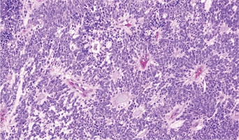
Medulloblastoma
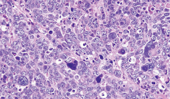
Medulloblastomas comprises the vast majority of pediatric embryonal tumors and by definition arise in the posterior fossa, where they constitute approximately 40% of all posterior fossa tumors. Other forms of embryonal tumors each make up 2% or less of all childhood brain tumors.The clinical feature
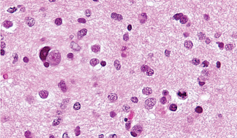
High-Grade Glioma
High-grade Gliomas (HGG) or astrocytomas in children nearly always result in a dismal prognosis. Although novel therapeutic approaches are currently in development, preclinical testing has been limited, due to a lack of pediatric-specific HGG preclinical models. These models are needed to help test
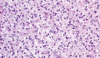
Low-Grade Glioma
Low-Grade Gliomas also called astrocytomas are the most common cancer of the central nervous system in children. They represent a heterogeneous group of tumors that can be discovered anywhere within the brain or spinal cord. Although surgical resection may be curative, up to 20% of children still su
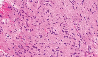
Diffuse Intrinsic Pontine Glioma
A presumptive diagnosis of DIPG based on classic imaging features, in the absence of a histologic diagnosis, has been routinely employed. Increasingly however, histologic confirmation is obtained for both entry into research studies and molecular characterization of the tumor.[5] New approaches with
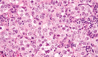
Germinoma
Germinomas are the most common type of CNS germ cell tumor and have a good prognosis.Germ cells are special types of cells that are present as the fetus (unborn baby) develops. These cells usually become sperm in the testicles or unfertilized eggs in the ovaries as the child matures. Most germ cell
Embryonal Tumor with Multilayered Rosettes
Nonmedulloblastoma embryonal tumors are fast-growing tumors that usually form in brain cells in the cerebrum, and most commonly occur in young children. The cerebrum is at the top of the head and is the largest part of the brain. The cerebrum controls thinking, learning, problem-solving, emotions, s
